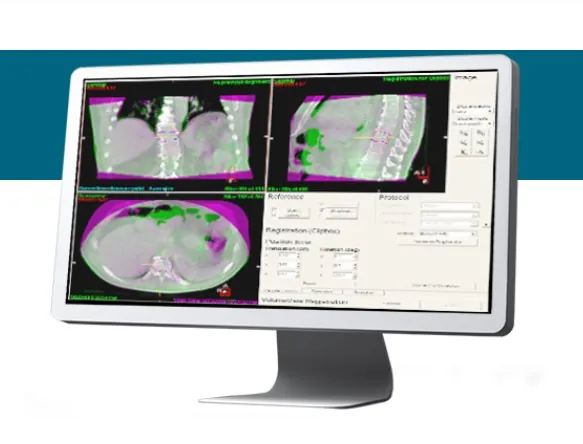
TREATMENT SOLUTIONS – ADVANCED IMAGING – XVI
XVI
2D, 3D and 4D kV imaging enables 2D and 3D volume imaging to
allow soft tissue detail to be seen in any area of the body immediately
prior to treatment.
2D, 3D and 4D soft tissue imaging
Tumor motion is a significant factor inhibiting targeting accuracy in
radiation therapy. The development of X-ray volume imaging (XVI)
and its suite of imaging tools was driven by the need to visualize
internal structures, within the reference frame of the treatment
system, to reduce geometric uncertainties. The ability to image at the
time of treatment with the patient in the treatment position reduces
issues relating to organ movement and inspires clinical confidence to
pursue advanced delivery techniques.
XVI imaging technology
Planning and treating in 3D requires verification in 3D. The 3D imaging
capability of XVI enables clinicians to take full advantage of complex
techniques without the need for implanted markers to visualize soft
tissue structures, target volume and the position of critical structures.
It allows precise registration of the reconstructed image data with the
historical CT planning data as a non-invasive procedure. XVI offers a
variety of image guided options to suit the individual needs of the
patient and the clinic XVI software offers the flexibility to vary the
dosage necessary to acquire a VolumeView image, depending on the
level of contrast required.
Clinical collaboration
Working closely with the Elekta Synergy Research Group, Elekta, in
collaboration with its clinical partners, became the first company to
support research on IGRT, the first to bring 3D volumetric imaging into
clinical use and the first to bring these solutions to the wider market.
Key advantages offered by VolumeView imaging at the time of
treatment include:
- Visualization of soft tissue dimensions and position
- Visualization of critical organs and tumors
- Visualization of bony anatomy and alignment in 3D
- Elimination of surrogate markers
- Ability to reduce the imaging dose
- Ability to overlay structures defined in treatment planning system
- Large field-of-view
- Simultaneous acquisition and reconstruction
Info Contact
| HEAD | OFFICE |
| Tel | +27 (11) 966 0600 |
| info@medhold.co.za | |
| Address | MSI Business Park, 68 Rigger Road, Spartan, Kempton Park, Gauteng, 1619 |


